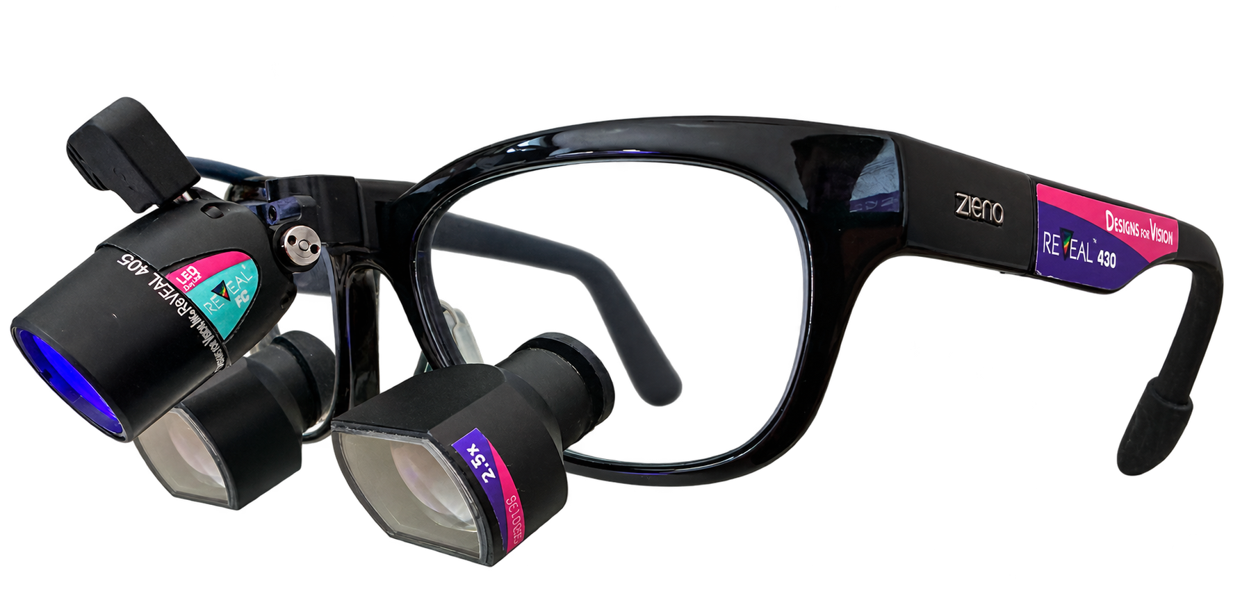
When white light alone offers little contrast
After an extraction, attention turns to the socket: which structures are directly visible, and can a retained tooth or root fragment be identified? Magnification helps you see small details. Another optical view can add to your assessment.
This is where REVEAL® comes in. Excitation light and matched viewing filters allow you to visualize fluorescence signals. You see these signals directly in the treatment area while both hands remain free for treatment.[1]
Why tooth and root tissue are of interest
Tooth root tissue has natural autofluorescence. The fluorescence of tooth and bone tissue can differ in intensity. This provides another view of the treatment area, where structures stand out differently than they do under white light.[2]
You assess these additional contrasts together with the shape and position of the visible structures. REVEAL® thus expands your view of the directly visible working area.
A look into clinical practice
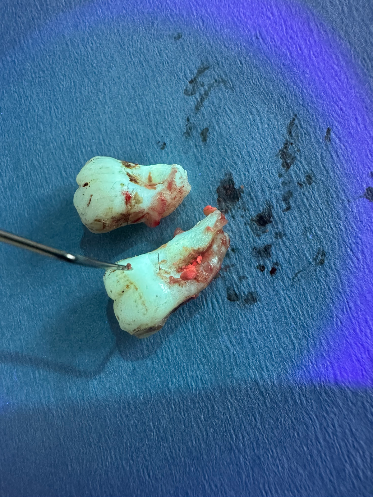
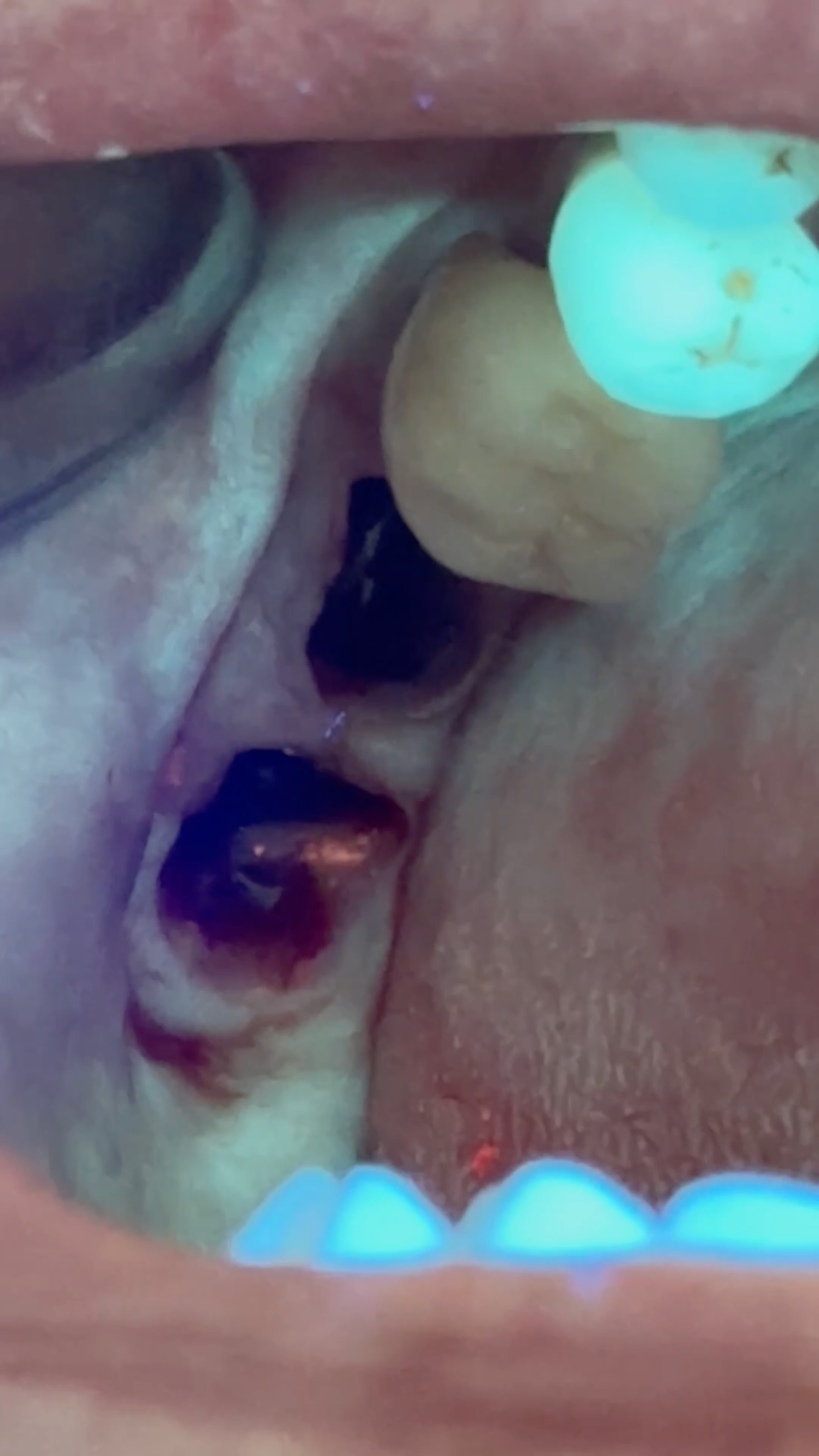
Tooth fragments and the extraction socket under REVEAL®
The following original images show two views of the same case after an extraction: the extracted tooth fragments and the socket. The bright tooth surfaces illustrate the additional fluorescence view.
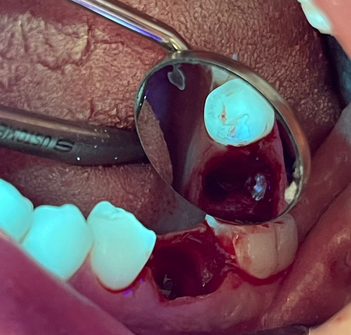
Searching for retained root fragments in a mirror view of the socket
A further clinical image shows a retained root fragment in a mirror view of the socket under REVEAL®. The additional fluorescence view is assessed together with shape, position and clinical findings.
17 seconds from clinical practice
Finding retained root fragments: Dr. Albrecht's experience
Dr. Florian Albrecht works with REVEAL® loupes from Designs for Vision with 3.0× magnification. In the video, he describes his personal experience of searching for retained root fragments.
He was skeptical at first. He now describes finding the small root fragment “relatively quickly” when he is able to dry the socket.
In further feedback, he sums up his experience as follows: “The endless searching is now a thing of the past.”
Why loupe optics from Designs for Vision matter
When searching an extraction socket, seeing fine detail and maintaining your working position go hand in hand. REVEAL® loupes combine magnification with matched fluorescence optics. Three points are particularly relevant to your choice:
-
View the detail under magnification
The loupe optics magnify details in the directly visible working area. Magnification and field of view are selected with you to suit your surgical tasks. Albrecht's 3.0× configuration is one specific clinician's example.
-
Match the focus to your working position
When custom-building your loupes, Designs for Vision takes into account working distance, declination angle and, where applicable, your prescription correction. This aligns the optics with the position in which you view the working area.[3]
-
Use magnification and fluorescence together
The excitation light and viewing filters work together. You see the additional information directly through the optics you are wearing. Both hands remain free for instruments and treatment.[1]
Your procedures. Your loupes.
What view do you need for your surgical tasks?
At Visiomedical, we discuss magnification, field of view and working distance with you, along with the additional view REVEAL® can provide. Tell us about your typical procedures and your previous experience with loupes.
Request a personal REVEAL consultationinfo@visiomedical.de