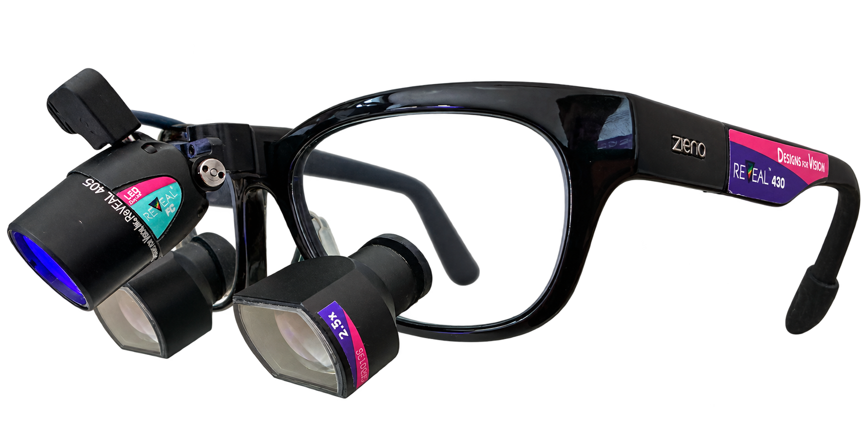
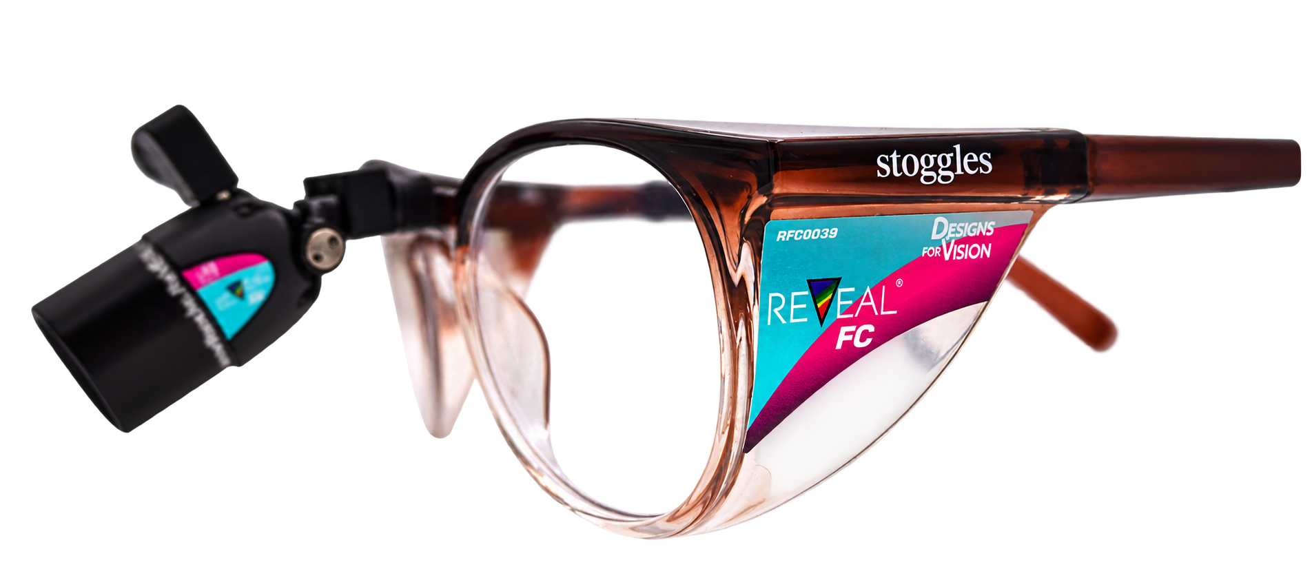
With the help of fluorescence, treat what others overlook.
An additional perspective on what you are treating. REVEAL FC makes fluorescence signals visible directly through matched filter optics – while leaving both hands free to work.
As custom-built loupes. Or as Stoggles without loupe magnification.
Three complementary images from the supplied implant dentistry case: a view of the operating field and two extraoral close-ups. The views illustrate the clinical context, not a guaranteed treatment outcome.
View of the operating fieldIntraoral fluorescence viewExtraoral close-up 01Additional view of the situationExtraoral close-up 02Additional view of the situation
The three views are not presented as a before-and-after comparison. Fluorescence color alone is not a clinical finding. Clinical images supplied for this draft; their source is separate from the publication images that follow.
BIO-OSS · COMPARING LIGHTING CONDITIONS
One material. Two lighting perspectives.
Geistlich Bio-Oss in clinical use: two original images from the Geistlich training presentation – without and with fluorescence illumination.
Both images reproduced unchanged. Slightly different viewing angles; no before-and-after comparison and no evidence of healing or complete decontamination.
01
PREVENTIVE DENTISTRY & CARIOLOGY
Deposits and tooth surface. With additional color information.
The lingual region shows different fluorescence patterns in deposits and an initial lesion described in the case report.
In everyday practice: Examine surfaces from an additional perspective and incorporate the visual impression into the clinical assessment.
Source & context
Hiltch, Steier & Poli de Figueiredo · 2024 · Case series involving three people, case 3 / Fig. 4. Different camera angles; not a before-and-after outcome.
A view into the cavity. Under two lighting conditions.
Treatment views from case 1 show white light and fluorescence during treatment of a carious tooth.
In everyday practice: Observe additional color information during instrumentation.
Source & context
Hiltch, Steier & Poli de Figueiredo · 2024 · Case series, case 1 / Fig. 2. The images show several treatment steps, not guaranteed treatment success. Clinical assessment and tactile criteria remain essential.
Case 2 combines fluorescence, white-light and radiographic views with images taken after opening the cavity.
In everyday practice: Use the fluorescence view as an adjunct – not as a substitute for necessary imaging.
Source & context
Hiltch, Steier & Poli de Figueiredo · 2024 · Case series, case 2 / Fig. 3. A case report does not establish general diagnostic superiority or reliable detection of deep lesions.
Deposits on a restoration. An additional way to see them.
An implant-supported restoration is shown under conventional light and with orange-red fluorescence signals.
In everyday practice: Examine accessible areas from an additional optical perspective – without inferring complete decontamination.
Source & context
Hwang, Blatz, Wolff & Steier · 2021 · Dentistry Journal 9(3):24, Fig. 2 · Review / concept paper, not a controlled efficacy study. Image taken with the system described in the article.
Four examples of different findings show fluorescence patterns on unremarkable surfaces, biofilm, demineralization and initial caries.
In everyday practice: An additional perspective on areas that are challenging to clean and assess visually.
Source & context
Bonfante et al. · 2025 · Dental Press Journal of Orthodontics · Clinical study with 60 participants. The study showed no statistically significant advantage for initial caries. The images are different examples, not a longitudinal sequence.
Fluorescence signals are not a diagnosis in their own right. Examination, necessary imaging and treatment decisions remain essential. The absence of a signal does not prove that biofilm is completely absent. Historical publication images document the systems investigated in each case, not every current REVEAL FC configuration.
ANOTHER APPLICATION · CRACKED TEETH
Examine crack structures under fluorescence illumination.
The clinical article “REVEAL: Cracked Teeth” presents optical approaches to tooth fractures. Some images also use sodium fluorescein – they are not all examples of purely dye-free autofluorescence.
Images show example configurations. We discuss availability, equipment, and costs with you. Our current offering does not combine ergonomic/Infinity VUE loupes with REVEAL.
04 / FROM INTEREST TO FIRST-HAND EXPERIENCE
Interesting in images. Crucial during the procedure.
We discuss whether REVEAL is right for you based on your needs. The initial consultation is free and without obligation.
Your work, previous experience with loupes and intended areas of use.
02
Arrange a suitable demonstration
Agree on demonstration options or a Stoggles in-office trial, including terms and availability.
03
Make an informed choice
Review the configuration, guidance on use, and personalized quote before deciding to buy.
05 / SCIENCE & CONTEXT
Know more. Make an informed decision.
Read the original articles on external servers. Study type and limitations are deliberately presented alongside the potential benefits in practice.
CARIOLOGY
Three clinical cases under different lighting conditions
The article combines fluorescence with clinical and radiographic interpretation. It illustrates applications but does not replace a robust comparative efficacy study.
Hiltch, Steier & Poli de Figueiredo · 2024 · Case series
Scientific background and a fluorescence-based approach to adjunctive visualization of deposits. A concept paper, not evidence of complete decontamination.
Biofilm, demineralization and caries around brackets
Clinical study with 60 participants on additional visual assessment. Results vary according to the findings; no general superiority across all applications.
Bonfante et al. · Dental Press Journal of Orthodontics · 2025
Crack structures and potential optical applications
An illustrative clinical article on adjunctive examination of tooth fractures; in part with additional sodium fluorescein. No general guarantee of diagnosis.
Krastev & Steier · Clinical Dentistry · August 2022 · Clinical article, from p. 87
Transparency matters: some authors disclose financial relationships with REVEAL technology. Study conditions and conflicts of interest can be found in the original articles. The image examples do not constitute an endorsement of Visiomedical by the authors.
06 / GOOD TO KNOW
Your questions. Clear answers.
What additional information can I see with REVEAL?+
You view fluorescence signals through the matched filter optics directly in the operating field. Patterns may vary depending on surfaces, deposits, tissues and materials. Their significance is assessed in the clinical context.
Does every red signal indicate the same finding?+
No. Color alone is not a diagnosis. Imaging conditions and the structure being examined must be taken into account. The additional view is assessed alongside the other findings.
Does REVEAL replace examination or necessary imaging?+
No. REVEAL is an adjunctive visualization aid. Examination, tactile criteria and necessary imaging remain part of your clinical assessment.
Does the absence of a fluorescence signal mean there is definitely no biofilm?+
No. The absence of a signal does not establish that biofilm is completely absent or that complete decontamination has been achieved. Optical feedback does not replace a complete clinical assessment.
Loupes or Stoggles – which are right for me?+
Loupes combine magnification tailored to you with a fluorescence view. Stoggles provide fluorescence visualization without loupe magnification or custom-built loupes. We discuss your requirements before you commit.
Can I try REVEAL in my practice?+
Demonstration options and a Stoggles in-office trial can be arranged personally. We discuss availability, guidance on use, and trial conditions in advance. The initial consultation is free; this does not mean that every in-office trial is free.
How much will my configuration cost?+
The optics, frame and equipment determine your personalized quote. We discuss costs transparently before you decide. A consultation inquiry is not an order.
THE NEXT STEP IS A CONVERSATION
Which optical technology suits your work?
Briefly describe your interest. We will discuss your options personally – free and without obligation.