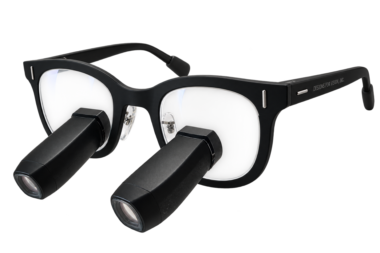
Why 3.5× should actually
magnify 3.5 times
A stated magnification is a technical claim. It should allow a meaningful comparison: how large does the same structure appear through the loupes compared with viewing it without magnification under defined reference conditions?
An independent laboratory study from 2013 shows that stated and measured values can differ. Nine medical loupe systems were tested for magnification and field of view, among other parameters. Some models labeled 2.5× achieved measured values between 2.0× and 2.7×. The results relate to the models available at that time; they are not a judgment on today's products. They do, however, illustrate why the figure alone is not a sufficient basis for comparison. Read the study by Neuhaus, Perrin and Lussi.
Designs for Vision calls its commitment to accurate magnification specifications “True Magnification”: the stated magnification level should correspond to the optical magnification actually delivered.
DESIGNS FOR VISION · TRUE MAGNIFICATION
True magnification.
Visible in comparison.
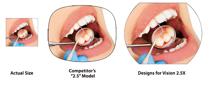
This is how Designs for Vision illustrates its commitment: the same clinical scene without magnification, through a competitor's model labeled “2.5×” and through its own 2.5× optics.
MANUFACTURER TEXT · ORIGINALLY TRANSLATED FROM ENGLISH
True magnification matters!
Designs for Vision has provided true magnification since 1961 and is accustomed to delivering the quality expected in dentistry. We believe that a 2.5× product should actually magnify the image to 2.5 times its size. Although some companies claim that magnification is not important, it is the most important feature in helping dental professionals see better. A loupe that magnifies the image only 2.0 times reduces the user's ability to visualize structures by 25 percent.
The purpose of magnification is to make an object appear larger. Designs for Vision offers accurate magnification with a clear image.
Distinguishing image size from detail resolution
Regarding the percentage in the manufacturer's text: using the same reference definition, a 2.5× image is 25 percent larger in width and height than a 2.0× image. Conversely, 2.0× produces an image that is 20 percent smaller than 2.5×. This does not demonstrate a corresponding gain or loss in discernible detail; image sharpness, contrast, lighting and vision also matter. The comparison shown is a manufacturer illustration, not a measurement of current competing products.
For you, this means choosing loupes based on clearly defined optical performance. A higher model number must not suggest greater magnification than the system actually delivers.
What diopters tell you –
and what they do not
Diopters and magnification factor describe different properties.
Diopters express the optical power of a lens. The magnification factor, by contrast, describes how large an object appears through an optical system compared with a defined reference view. For simple magnifiers, a relationship between optical power and magnification can be calculated under specified conditions. Medical loupes, however, consist of several optical elements. Their magnification therefore cannot be derived from a single diopter value using a general conversion formula. Optical fundamentals: MIT, University of Tennessee.
The absence of a diopter specification is therefore not proof of an unsupported magnification claim. Conversely, a diopter value alone does not confirm a particular magnification factor.
The key question is: can the supplier explain what the stated magnification means and how it was determined? Without this explanation, you are missing an essential basis for comparison.
What difference does
higher magnification make?
Using the same reference basis, the difference between 2.5× and 3.5× is clear: the factor increases by 40 percent. The same structure therefore appears 40 percent larger in both width and height.

However, this does not automatically mean that you can discern 40 percent more detail. Image sharpness, contrast, lighting and your own vision also matter.
Magnification therefore answers one question first: how large does the detail appear? How well you can assess it depends on the quality and fit of the entire system.
Five questions
for a meaningful comparison
- What does the stated magnification mean?Ask how the value was determined and which reference conditions apply. A model designation is no substitute for this information.
- At what working distance do the specifications apply?Magnification and field of view should be considered together with the intended working distance. Compare under the same conditions wherever possible.
- How much of the surrounding area remains visible at the desired magnification?Check whether you can see enough of the surrounding area as well as the detail itself to guide your instruments safely and orient yourself in the working area.
- Is the image in focus in my natural working position?Try the loupes in your usual working posture. Working distance, viewing angle, optical alignment and, where applicable, prescription correction must suit you.
- How well can I see the same fine structures?For a direct comparison, use the same suitable test object and, wherever possible, the same lighting. Assess image size, sharpness and contrast at the edge of the field of view as well. This hands-on comparison helps you choose; an exact magnification measurement requires an appropriate measurement setup.
A reliable figure
is the starting point for a good decision
The right loupes are those that allow you to see the necessary details and work precisely in a comfortable working position. The highest magnification alone does not meet this requirement.
The figure must be accurate. Field of view, image quality, working distance and custom fitting must also work together. These factors make a magnification level a sound choice for your everyday clinical practice.
At Visiomedical, this is the starting point for our advice on loupes from Designs for Vision: together, we establish which magnification your tasks require and which equipment fits the way you work.
Free, no-obligation advice from Visiomedical.